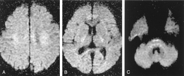
Fig 6.
In b = 2000 images, densely packed white matter tracts normally shows relative hyperintensities compared with gray matter, especially at the corona radiata (A), internal capsule (B), and the lower pons level (C). These normal relative hyperintensities of white matter tracts should not be erroneously diagnosed as ischemic lesions.

