Fig 2.
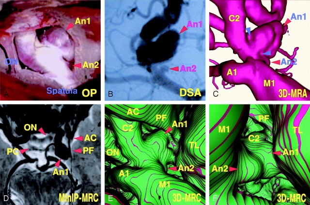
Case 1, an 83-year-old woman with a large unruptured right internal carotid–posterior communicating artery aneurysm and a small anterior choroidal artery aneurysm.
A, Operative photograph showing the caudal view of the right superolateral aspect of the posterior communicating artery aneurysm (An 1), the anterior choroidal artery aneurysm (An 2), and the right optic nerve (ON).
B, Digital subtraction angiogram, right oblique projection, showing the aneurysms (An 1, An 2).
C, 3D MR angiogram, similar projection to the operative view in panel A, showing the arterial components of the aneurysmal complex. Note the slightly concave surface at the neck (arrowheads).
D, MinIP image obtained from MR cisternography, superoinferior projection, showing the cisternal structures with negative shadows in contrast to the surrounding CSF with a positive shadow. ON, optic nerve; AC, anterior clinoid process; PC, posterior clinoid process; PF, petroclinoid dural fold.
E, Conventional 3D MR cisternogram, similar projection to the operative view in panel A, depicting the contours of the aneurysmal complex (An 1, An 2, A1, C2, M1), the right optic nerve (ON), the anterior clinoid process (AC), the petroclinoidal dural fold (PF), and the temporal lobe (TL).
F, Conventional 3D MR cisternogram, virtual viewpoint from superoposterior projection, showing the aneurysmal complex and perianeurysmal environment.

