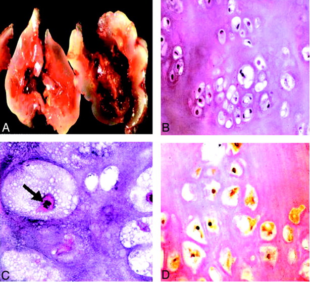
Fig 2.
Histopathologic findings.
A, Gross specimen shows a bluish white, pearly, glistening lobulated tumor.
B, Low power slide shows a hyaline tumor of low to moderate cellularity with mild variation in size and shape of the tumor cells with mostly small and dark nuclei, showing mild nuclear pleomorphism (hematoxylin and eosin; original magnification, ×40).
C, High power slide reveals mitotic figures (arrow). Findings consistent with the appearance of a conventional chondrosarcoma (hematoxylin and eosin; original magnification, ×200).
D, Nuclear and, to a lesser degree, cytoplasmic immunoreactivity for S-100 protein are present (immunoperoxidase; original magnification, ×100), indicative of a cartilaginous tumor.

