Fig 2.

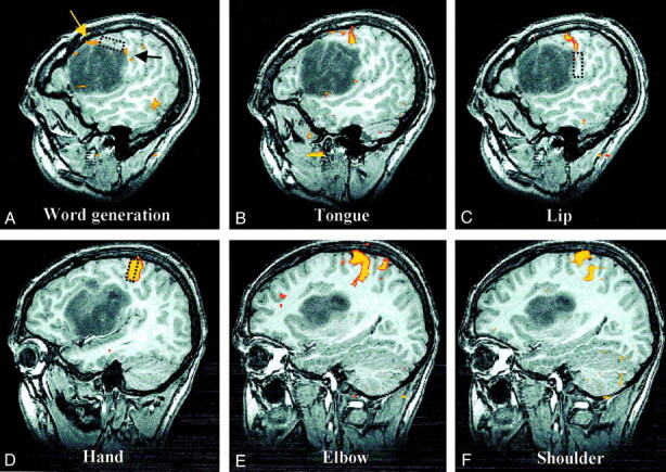
Sagittal spoiled gradient-echo and fMR images of the left hemisphere show cortical activation, compared with intraoperative mapping sites.
A, Language activation. BOLD language activation is displaced superiorly (yellow arrow) but located immediately anterior to language function mapped intraoperatively (rectangle). A small amount of additional activation associated with the word-generation task (black arrow) corresponds more closely to the intraoperative location of lip function (see image in C).
B, Tongue activation. BOLD tongue activation is situated between intraoperative maps of lip and language function, as would be expected, but intraoperative attempts to find tongue motor function were unsuccessful.
C, Lip activation. Corticonuclear (tongue and lip) BOLD activation is in superiorly displaced precentral gyrus at the superior-posterior edge of the tumor located immediately above the area of lip function mapped intraoperatively (rectangle).
D, Hand activation. The corticospinal (hand, elbow, and shoulder) cortical function is in the normal portion of the precentral gyrus superior and posterior to the tumor. With these three areas, only intraoperative mapping of hand function was attempted, and the result corresponded precisely with the location of BOLD activation (rectangle).
E, Elbow activation.
F, Shoulder activation.
