
Fig 3.
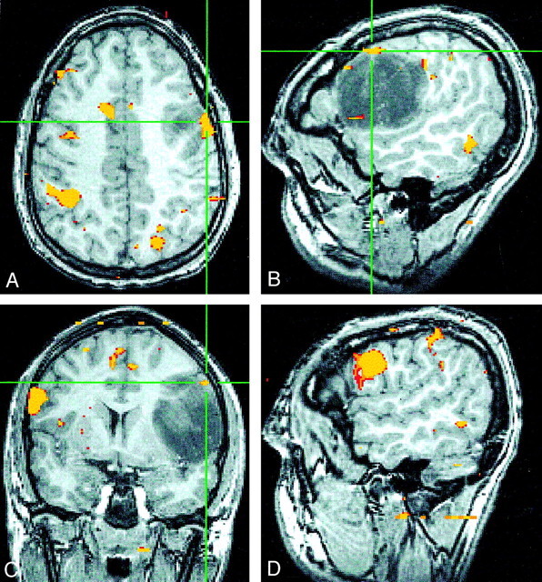
Images falsely suggesting strong right-hemisphere dominance.
A–C, Axial (A), sagittal (B), and coronal (C) SPGR fMR images show minimal left-frontal language activity (crosshairs) during a word-generation task.
C and D, Coronal (C) and sagittal (D) fMR images show robust activity in the right IFG and inferior frontal sulcus that falsely implies right-hemisphere language dominance in this individual.