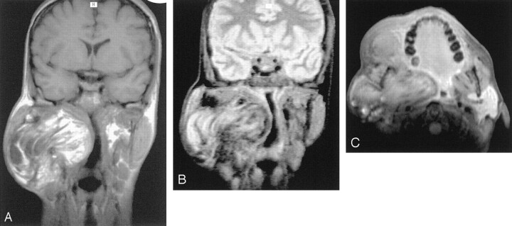
Fig 3.
MR images obtained with a 1.0-T superconductive imaging unit.
A, Coronal spin-echo T1-weighted MR image (512/17) shows an inhomogeneous hyperintense lesion located in the parotidomasseteric region and extending deeply toward the parapharyngeal space.
B, On spin-echo T2-weighted MR image without fat suppression (2300/80), decreasing signal intensity is appreciable and irregular linear regions of markedly hypointense signal are present.
C, Axial contrast-enhanced fat-suppressed spin-echo T1-weighted MR image (900/22) shows partial suppression of the lesion, consistent with fat. The lesion nevertheless shows ill-defined margins without a clear plane of cleavage between the mass and the adjacent muscular structures, especially the pterygoid and masseter muscles that appeared displaced and compressed. No evidence of contrast enhancement within the mass can be seen.

