
Fig 5.
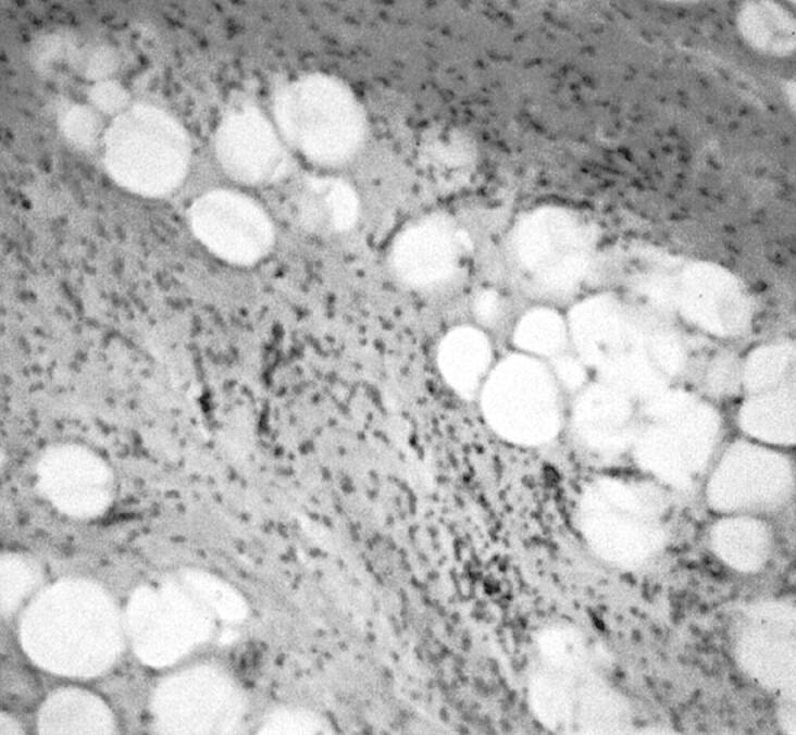
Pathologic specimen (hematoxylin and eosin; original magnification, ×400). The lesion is composed of uniform-appearing mature adipocytes, with the presence of myxoid hypocellular areas and infiltrates of inflammatory cells, especially lymphocytes. Diffuse infiltration of entrapped striated muscles can be seen; no evidence of lipoblasts, cellular atypism, or mitoses indicating liposarcoma is seen.