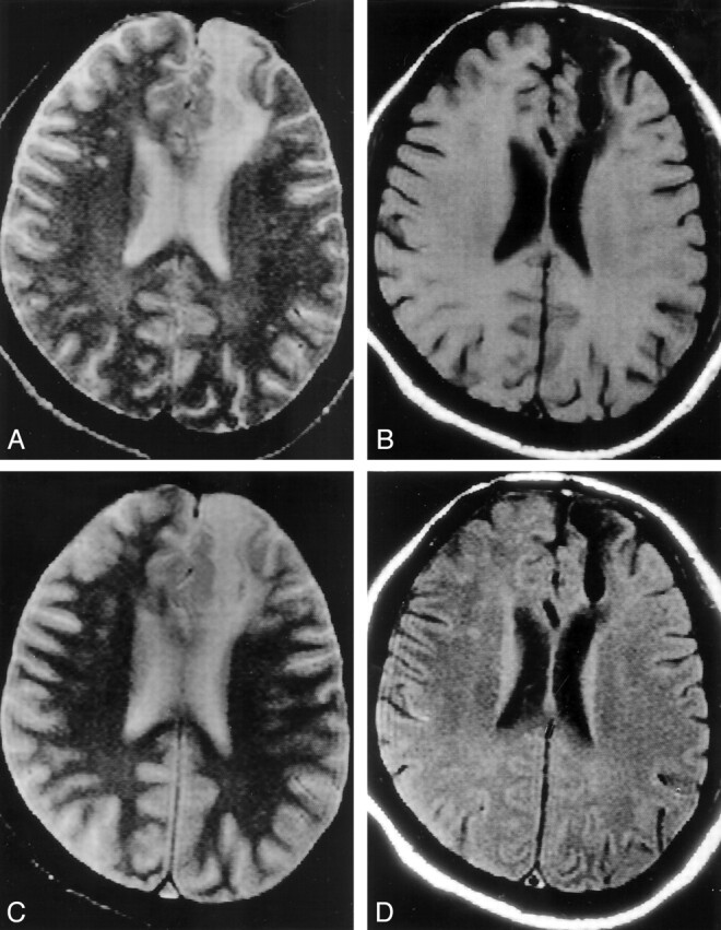
Fig 1.

Patient without seizures after head trauma (disease control).
A and B, Axial T2-weighted image (A) through the lateral ventricles shows a large hyperintensity involving the left frontal region; this area appears hypointense on the T1-weighted image (B).
C, T2*-weighted image does not show any bloom effect to suggest hemosiderin deposit.
D, T1-weighted MT image does not show abnormality beyond the abnormality seen on the T2-weighted (A) and T1-weighted (B) images.
