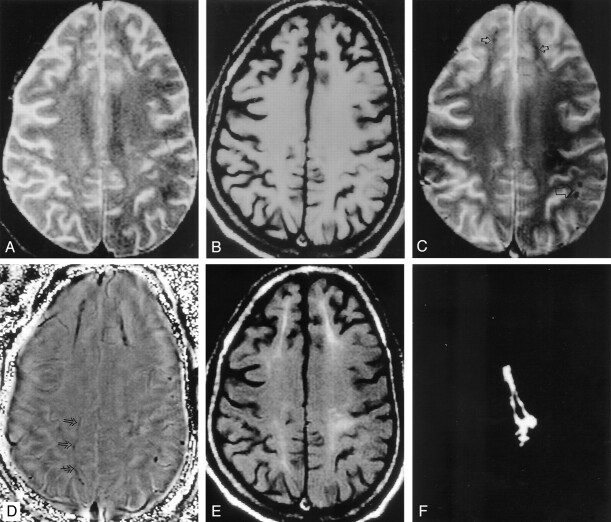
Fig 3.
Gliosis around the hemosiderin deposit in the brain parenchyma of a patient with intractable PTE.
A and B, Axial T2-weighted image (A) through the supraventricular region shows subtle hyperintensity in the white matter involving both sides of the frontal lobe and the left parietal region; this is not visible on the corresponding T1-weighted image (B).
C, T2*-weighted image shows multiple hypointense focal round lesions (large arrow) in the left parieto-occipital region and linear hypointensities (small arrows) in both frontal regions and the left parietal region.
D, In addition, phase-corrected GRE image shows subtle hypointensity (arrows) in the right parasagittal region that was not visible in C. All these hypointensities that are seen as negative signal intensity suggest hemosiderin deposition.
E, Corresponding T1-weighted MT image shows high signal intensity around the linear hemosiderin deposits, which is suggestive of gliosis.
F, Difference of the segmentation of the abnormality seen on the phase-corrected GRE image and T1-weighted MT image in the left frontal region confirms that the MT abnormality is beyond the hemosiderin deposit. The central black region represents the hemosiderin seen on the phase image.

