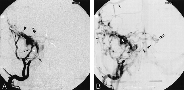
Fig 2.
Dural arteriovenous fistula of the lesser sphenoid wing.
A, Early filling of a selective right external carotid artery injection in the anteroposterior projection. Nourishing branches from the right middle meningeal artery (black arrows) feeds a dilated superficial middle cerebral vein (black arrowhead). The superficial middle cerebral vein drains into a laterocavernous sinus (white arrowhead), which is also directly involved in the dural arteriovenous fistula and is fed by branches of the right middle meningeal artery. Early anterograde filling of the cavernous sinus is observed. There is retrograde filling into the right basal vein (white arrows) and into the right peduncular vein, through the right deep middle cerebral vein.
B, Selective right external carotid injection, later in the venous phase. Retrograde filling of the right superficial cortical veins (black arrow) via the right superficial middle cerebral vein is now observed. The left basal vein (black double arrow) is now also opacified via the peduncular veins, and the straight sinus is clearly delineated. There is infratentorial drainage of the fistula into the anterior and lateral pontomesencephalic veins (black arrowheads). The saccular dilatation of the superficial middle cerebral vein is clearly visible (asterisk).

