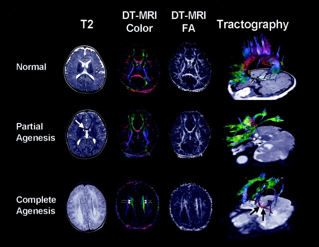
Fig 1.
Anatomic T2-weighted images, diffusion tensor-based color maps, FA maps, and FT of callosal dysgenesis. Upper row depicts normal findings of corpus callosum; i.e., clear red fibers and high FA bundles crossing midline at diffusion tensor MR imaging. Tractography shows interhemispheric fiber connections through corpus callosum; i.e., red-colored fibers in the midline. Middle row shows partially developed corpus callosum in genu portion (white arrow). Tractography demonstrates fibers from parieto-occipital regions converging into small red-colored genu as well as fibers from frontal lobes. Fibers from the posterior part run anteriorly and form longitudinal green-colored fibers, the Probst bundle. In complete agenesis of corpus callosum, the Probst bundle is more apparent as thick, green, high-FA fibers medial to lateral ventricle (double arrows). Tractography shows thick bundles of green color consist of various fibers from ipsilateral hemisphere, Probst bundle, and thick red-colored AC (black arrows).

