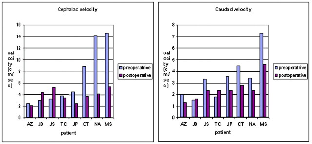
Fig 1.
Graph displaying the peak caudad and cephalad velocities (in cm/s) in the subarachnoid space of the foramen magnum in patients with a Chiari I malformation before and after suboccipital decompression. In most patients, the caudad (systolic) and cephalad (diastolic) velocities are diminished following surgery.

