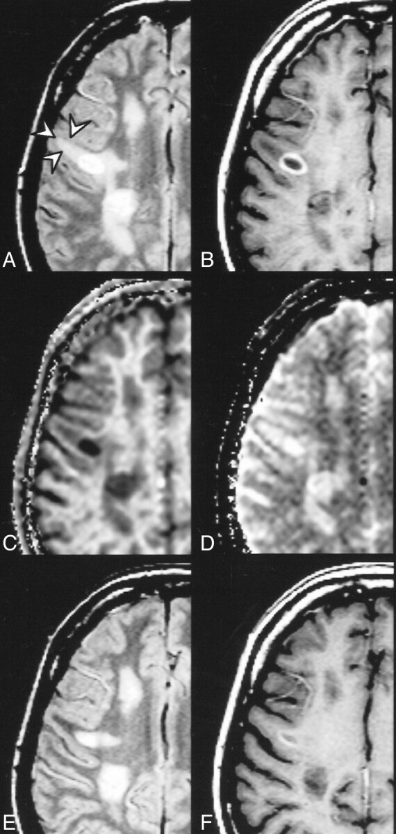
fig 4.

Ring-enhancing lesion surrounded by edema (arrows) and multiple non-active lesions. Proton density–weighted (2060/20/1) (A), postcontrast T1-weighted (510/14/2) (B), kfor (C), and T1free (D) images. The quantitative analysis in the acute lesions yielded the following results: ring-enhancing lesion: MTR = 16.4 %, kfor = 0.13 s−1, T1free = 1.5 s; edema: MTR = 41.1 %, kfor = 0.56 s−1, T1free = 1.2 s. Proton density–weighted (E) and postcontrast T1-weighted (F) images acquired 6 weeks later. Disappearance of perifocal signal abnormality supports the assumption of edema
