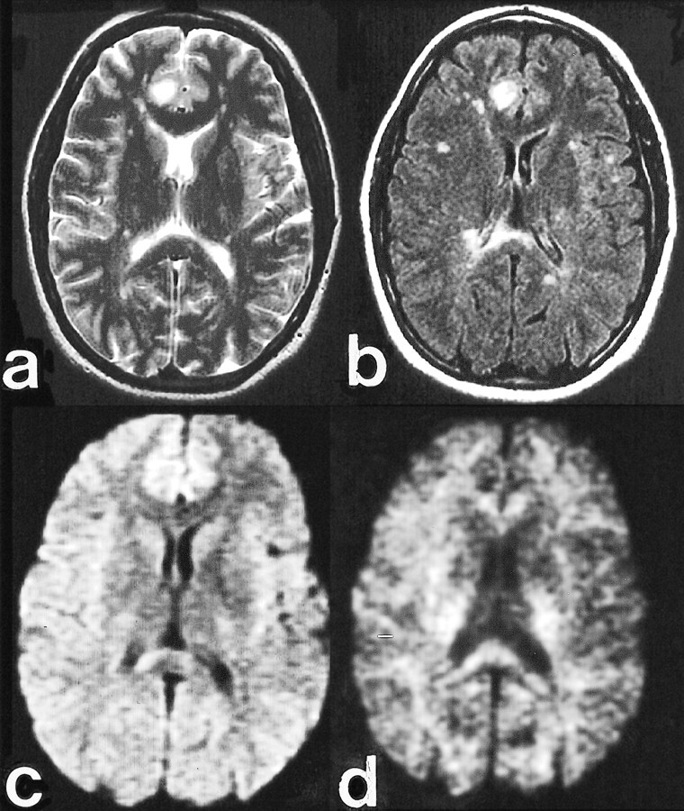
fig 4.

Clinical case of multiple sclerosis.
A, Fast spin-echo axial T2-weighted image (4000/102/2 [TR/TE/excitations]) shows a dominant subcortical right cingulate gyrus lesion with marked hyperintensity.
B, Fast-FLAIR image (10000/145/2200/1 [TR/TE/excitations]) highlights additional subcortical and periventricular plaques, including a large lesion in the right side of the splenium.
C, Diffusion-weighted image (10000/72/1) obtained with b = 1000 s/mm2 reveals slight hyperintensity in the right cingulate lesion, similar to the contralateral mesial frontal region, and hyperintensity in the right splenium lesion.
D, Diffusion-weighted image (10000/96/1) obtained with b = 3500 s/mm2 reveals marked hypointensity in the right cingulate lesion, likely reflecting reduced T2 weighting and less T2 shine-through. Persistent hyperintensity in the right splenium lesion is likely due to the normal anisotropy of the splenium and inconspicuity of hypointense lesions adjacent to the CSF spaces. This emphasizes the complementary relationship of the diffusion-weighted acquisitions obtained at standard and high b values.
