fig 1.

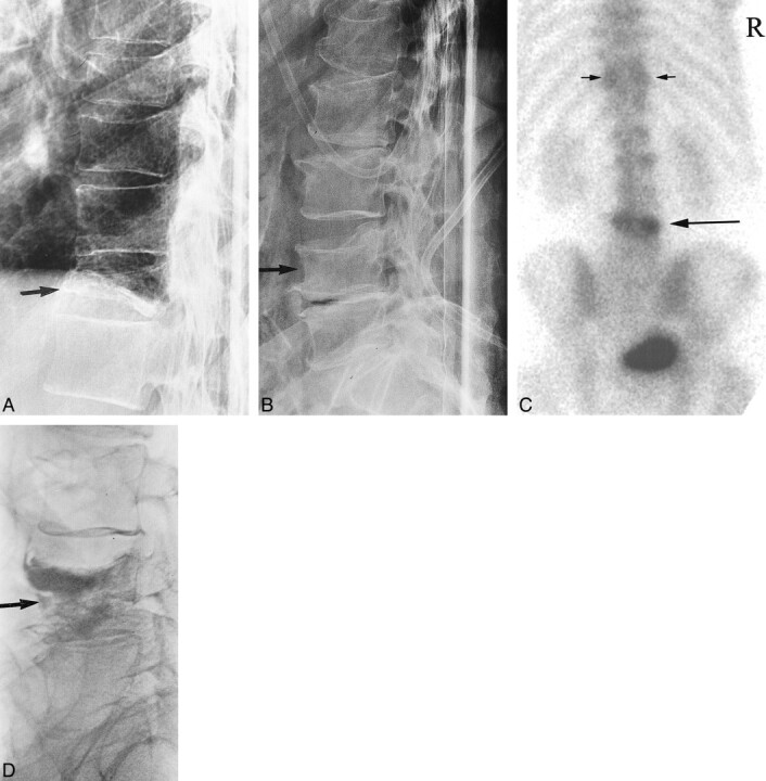
Images from the case of a 79-year-old man who presented with a history of back pain of several months' duration. Although the patient complained of mid and low back pain, he did not experience localizing pain during physical examination.
A, Lateral view plain film radiograph of the thoracic spine shows wedge compression fracture at T11 (arrow).
B, Lateral view plain film radiograph of the lumbar spine shows wedge compression fractures at L4 (arrow) and endplate compression fractures of L1, L2, and L3.
C, Posterior planar Tc99m-MDP bone scan image shows markedly increased activity at L4 (large arrow) and minimal increased activity over the pedicles or facets of T11 (small arrows). Based on increased activity over L4 and relative lack of increased activity at other fracture sites, we proceeded with a single-level vertebroplasty at L4.
D, Lateral view plain film radiograph of the lumbar spine after vertebroplasty shows barium-opacified methylmethacrylate within the L4 vertebral body (arrow). The patient experienced complete pain relief after treatment. Before treatment, the patient's mobility was limited to walking with assistance. After treatment, he was able to walk without assistance.
