fig 2.

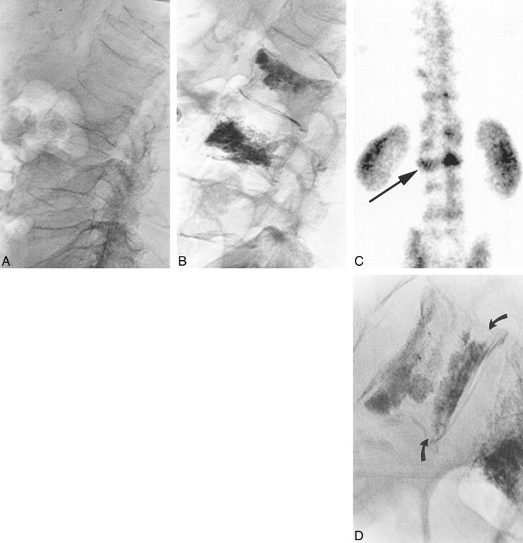
Images from the case of a 77-year-old man who presented with back pain of 3 months' duration.
A, Lateral view plain film radiograph obtained over the lumbar spine shows fractures at L1–L5. No previous images were available to date the onset of fracture at any level. However, because the patient experienced focal tenderness over the spinous processes of L2 and L3 without tenderness over the other fracture levels, we elected to proceed with vertebroplasty at L2 and L3, without performing preprocedural bone scan imaging.
B, Postprocedural lateral view plain film radiograph obtained over the lumbar spine shows barium-opacified methylmethacrylate within the L2 and L3 vertebral bodies. Cement injection at L3 traversed the entire marrow cavity. Cement selectively traversed the superior endplate region at L2, which is commonly seen as the cement naturally extends into fracture lines. Subjectively, the patient improved “about 50 percent,” but he requested further treatment, if possible. Bone scan imaging was performed because of uncertainty regarding further therapy.
C, Posterior planar Tc99m-MDP2 bone scan shows increased activity in the region of the L2 fracture (arrow). Based on the bone scan image, which suggested ongoing bone turnover in L2, we selectively targeted the inferior aspect of L2 for the second vertebroplasty treatment.
D, Postprocedural lateral view plain film radiograph shows barium-opacified methylmethacrylate within the inferior aspect of L2 (curved arrows). The patient noted complete pain relief after the second vertebroplasty at L2, stating that he felt “the best he has felt in years.”
