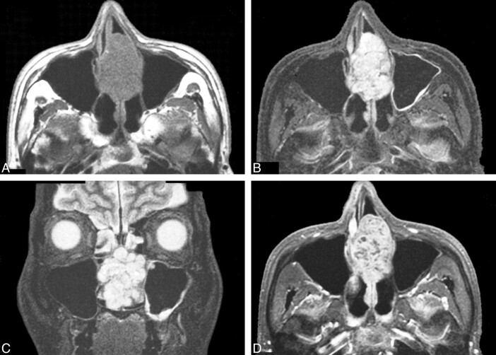
fig 2.
MR images of the intranasal mass.
A, Axial T1-weighted image (500/14/2 [TR/TE/excitations]) shows the tumor has destroyed the nasal septum and occupies both nasal cavities. The tumor is isointense to brain.
B, Axial fast-STIR image (4000/30/2) shows a lobular, high signal intensity tumor. The tumor abuts the nasal septum to the right and has destroyed it. The left maxillary sinus is intact.
C, Coronal fast-STIR image (4000/30/2) also shows a well-marginated, lobular, high-signal-intensity mass occupying the midline nasal cavity. The skull base is intact.
D, Axial contrast-enhanced fat-suppressed T1-weighted spin-echo image (400/20/2) shows marked enhancement with foci of unenhanced lower signal intensity within the body of the tumor.

