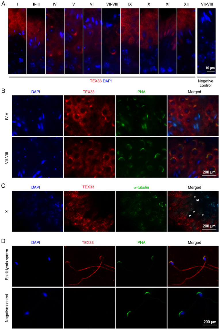
Figure 2.
Localization of TEX33 during spermatogenesis. (A) Immunofluorescence staining of TEX33 in the seminiferous tubules during different stages of spermatogenic development (stages I–XII). (B) Co-immunofluorescence staining of TEX33 with PNA-FITC in the seminiferous tubules during different stages of spermatogenic development. (C) Co-immunofluorescence staining of TEX33 and α-tubulin in elongating spermatids during spermatogenic stage X. (D) Co-immunofluorescence staining of TEX33 with PNA-FITC in sperm. TEX33, testis-expressed protein 33; PNA-FITC, FITC-conjugated peanut agglutinin; DAPI, 4,6-diamidino-2-phenylindole; M, manchette (indicated by arrowheads).

