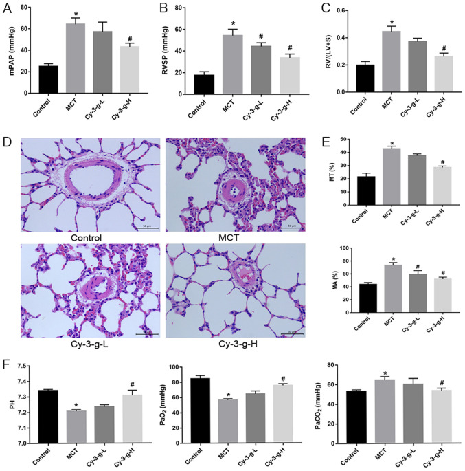
Figure 1.
Protective effect of Cy-3-g on hemodynamics in rats with pulmonary artery hypertension induced by MCT. Rats were randomly assigned into four groups (n=15): Control group, model group (MCT), Cy-3-g-L group and Cy-3-g-H group. On the first day of the experiments, rats in the MCT, Cy-3-g-L and Cy-3-g-H groups were injected with MCT (60 mg/kg). On days 2–28, the rats in the Cy-3-g-L and Cy-3-g-H groups received a gavage of 200 and 400 mg/kg body weight, respectively. Changes in: (A) Mean mPAP, (B) RVSP and (C) RV hypertrophy index. (D) Representative images of hematoxylin and eosin staining of lung tissue. (E) Quantitative data of percentage of MT and MA of the pulmonary artery. (F) Changes in pH and partial pressure of PaO2 and PaCO2. Data are shown as the mean ± SD. *P<0.05 vs. control; #P<0.05 vs. MCT. MCT, monocrotaline; Cy-3-g, cyanidin-3-O-β-glucoside; L, low-dose; H, high-dose; mPAP, pulmonary artery pressure; RVSP, right ventricular systolic pressure; RV, right ventricular; MT, medial wall thickness; MA, medial wall area; PaO2, arterial oxygen; PaCO2, arterial carbon dioxide.

