fig 1.
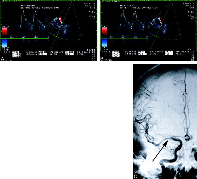
Comparison between angle-corrected and uncorrected flow velocities and angiographic findings in a 54-year-old woman with MCA stenosis.
A and B, At TCCD sonography, the sample volume is placed approximately 10 mm from the internal carotid artery bifurcation and within the color image of the right MCA. The velocity spectrum is adjacent to the left of the image of the artery. The uncorrected peak systolic velocity is 149 cm/s (B).
C, Angiogram shows a lesion causing a stenosis of more than 50% (arrow) in the right MCA M1 segment. The angle-corrected peak systolic velocity is 204 cm/s.

