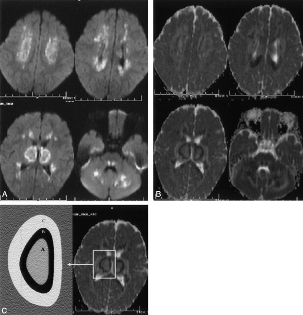
Fig 2.
Diffusion findings in acute-stage ANE in a 2-year-old girl. A, Diffusion-weighted MR images (b = 1000) show bilateral symmetric diffuse hyperintense lesions in the cerebellar-cerebral white matter, thalami, and pontine tegmenta. B, The ADC map shows three different patterns of the thalamus and cerebral white matter. The center of the lesions shows higher ADC values than those of normal parenchyma, the peripheral portion of central lesions shows very low ADC values, and outside the thalamus and cerebral white matter are high ADC values, findings compatible with vasogenic edema. Note that the splenium of corpus callosum, pontine tegmanta, optic radiatia, and cerebellar white matter have low ADC values in the central portion and high ADC values in the peripheral portion. C, Mean ADC values and pattern on the right thalamus (A, center of thalamic lesions; B, periphery of the central thalamic lesions; C, outside portions of the thalamic lesions).

