
Fig 3.
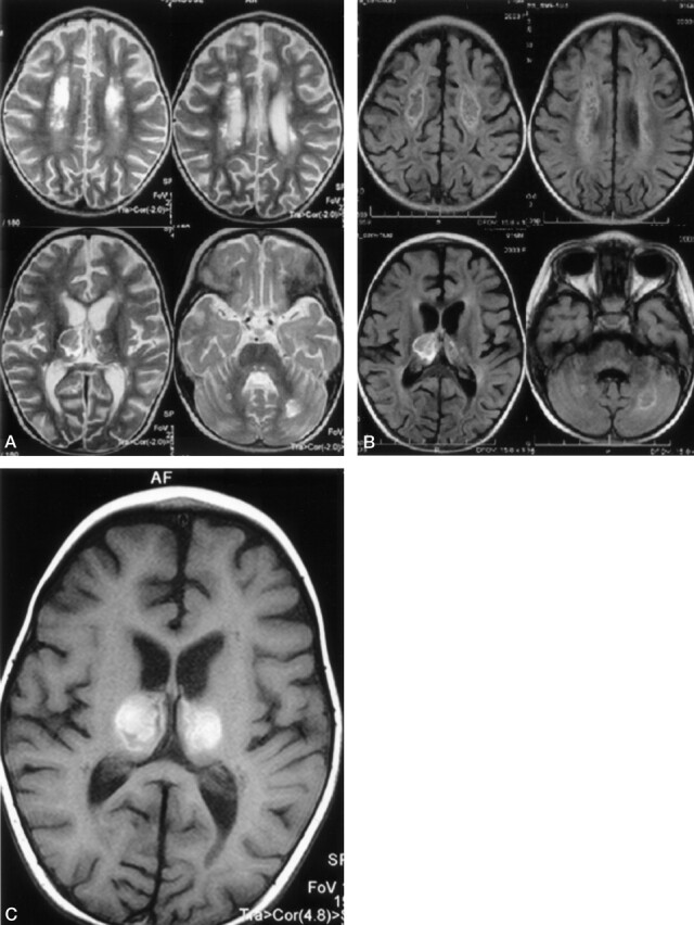
On follow-up MR examination, 25 days after onset of symptoms, T2-weighted (A) and fluid-attenuated inversion recovery (B) MR images of brain show neuronal tissue destruction on the central portion of cerebral white matter, bilateral optic radiation, deep cerebellar white matter, and pontine tegmanta. Necroses are also seen on the central portion of thalamus and deep cerebral white matter. T1-weighted MR image (C) shows subacute hemorrhagic changes in the central necrotic portion of thalamus.