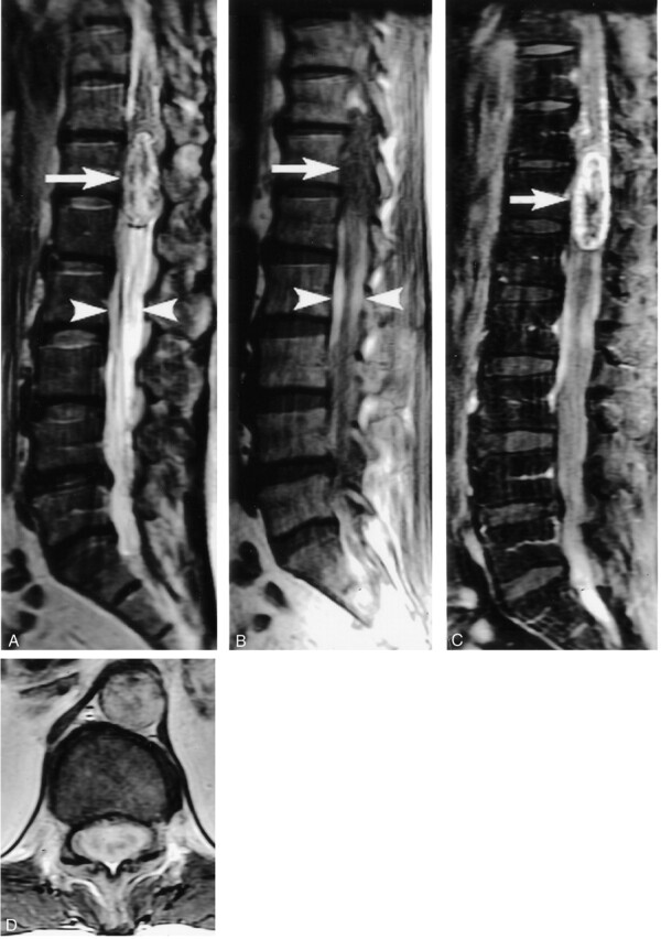
Fig 2.

Thoraco-lumbar spinal tumor. Sagittal T2- (A) and T1-weighted images (B) show a large mass at the conus medullaris with heterogeneous signal intensity (arrows). The two hyperintense SHDs may be seen located caudal to the mass (arrowheads).
After intravenous contrast material injection (C and D), there is heterogeneous enhancement of the mass.
