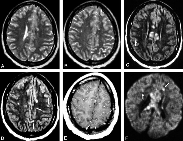
Fig 1.
Patient 1. Initial MR examination at 2 weeks from the onset of symptoms.
A and B, Axial T2-weighted images show multiple hyperintense lesions involving the corpus callosum and cerebral white matter.
C and D, Axial FLAIR images at almost the same levels as in A and B show hyperintense lesions not only in the corpus callosum and cerebral white matter but also in the cerebral cortex (arrows).
E, Contrast-enhanced axial T1-weighted image shows diffuse leptomeningeal enhancement.
F, Axial DWI shows several hyperintense lesions in the cerebral white matter (arrows), with a cluster of lesions in the corpus callosum.

