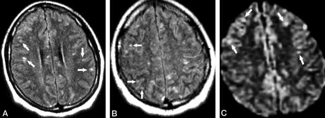
Fig 2.
Patient 1. Second MR examination shortly after relapse of the symptoms (8 weeks from initial onset).
A and B, Axial FLAIR images show that the lesions in the corpus callosum have decreased in size and number compared with those in Figure 1. However, new punctate hyperintense lesions appeared in the cerebral cortex (arrows).
C, Axial DWI also reveals the scattered hyperintense lesions in the cerebral cortex (arrows).

