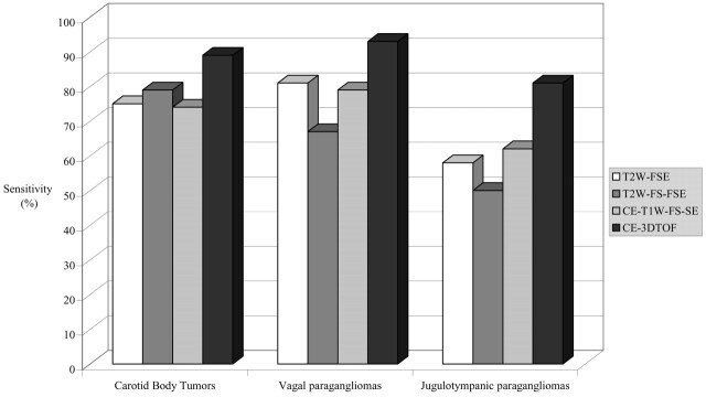
Fig 1.
Bar graph shows sensitivity (mean for both observers) for detection of carotid body tumors, vagal paragangliomas, and jugulotympanic paragangliomas for each MR imaging technique. T2W-FSE, T2-weighted fast spin-echo imaging; T2W-FS-FSE, T2-weighted fat-suppressed fast spin-echo imaging; CE-T1W-FS-SE, contrast-enhanced T1-weighted fat-suppressed spin-echo imaging; CE-3DTOF, contrast-enhanced 3D time-of-flight imaging.

