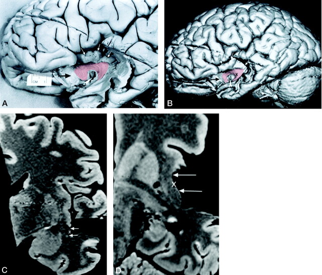
Fig 3.
Method used to identify the location of the dissected surface of the uncinate fasciculus on reformatted cross-sectional MR images.
A, Photograph of the lateral surface of the dissected brain specimen. Branches of the middle cerebral artery have been dissected away, leaving the horizontal segment (M1) of the middle cerebral artery as a landmark (arrowhead). The lower part of the cortex of the insular gyri (white arrows) and the posterior orbital gyrus (black arrow) have also been removed by dissection. A segment of the surface of the uncinate fasciculus (transparent red) is visible and courses from the temporal lobe into the white matter of the insula. It passes anteriorly around the middle cerebral artery and into the posterior orbital gyrus. To demonstrate the underlying dissected uncinate fasciculus, we used a 10% transparent red that results in a pink appearance.
B, 3D MR rendering of the dissected specimen in A demonstrates the accurate surface anatomy that can be depicted in this way. A segment of the surface of the uncinate fasciculus is color coded transparent red. The middle cerebral artery (arrowhead), the cortex of the insular gyri (white arrows), and the posterior orbital gyrus (black arrow) can be identified.
C and D, Coronal (C) and axial (D) reformatted cross-sectional MR images show the location of the dissected segment of the surface of the uncinate fasciculus (arrows). These cross-sectional images were generated at a level indicated by a cursor (X) placed on the surface of the uncinate fasciculus on the 3D MR rendering in B. The position of the cursor on the cross-sectional images indicates the surface of the uncinate fasciculus. Coregistration of the cursor permitted comparison of localization on the dissected surface with corresponding coronal and axial MR images.

