fig 1.

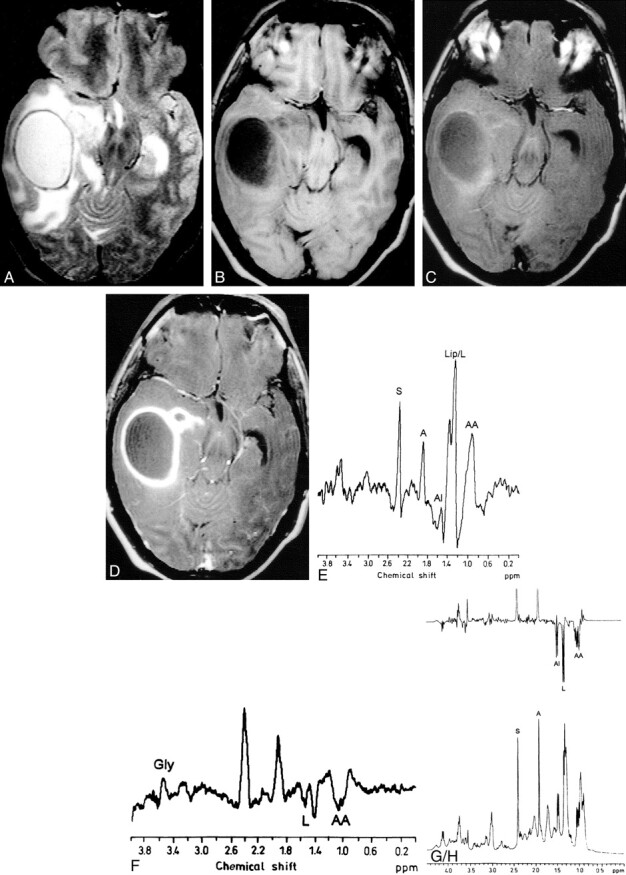
Pyogenic brain abscess.
A, T2-weighted MR image through the temporal lobe shows a well-defined hyperintense lesion in the right temporal lobe with peripheral hypointense rim, perifocal edema, and mass effect on the ventricular system.
B, T1-weighted image shows the lesion as hypointense with isointense wall.
C, MT T1-weighted image shows minimal hyperintesity. The MT ratio from the wall is 31.25.
D, Postcontrast MT T1-weighted image shows enhancement of the rim of the lesion.
E and F, In vivo MR spectra obtained using STEAM (E) at 3000/20/30/128 (TR/TE/TM/excitations) and spin-echo (F) at 3000/135/128 (TR/TE/excitations) show presence of glycine (Gly) at 3.56 ppm; succinate (S) at 2.4 ppm; acetate (A) at 1.92 ppm; alanine (Al) at 1.5 ppm; lipid/lactate (Lip/L) at 1.3 ppm; and leucine, isoleucine, and valine (AA) at 0.9 ppm.
G and H, Ex vivo MR spectroscopy with single-pulse (G) and spin-echo (H) imaging confirm the assignments seen in vivo.
