fig 1.

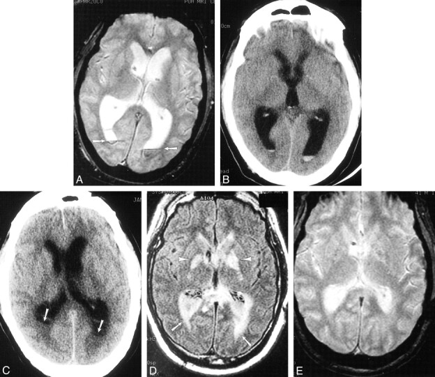
Case 3. 41-year-old patient who underwent routine postoperative MR imaging 4 days after resection of a posterior fossa ependymoma. Enterobacter aerogenes grew on culture of ventricular CSF.
A, Axial gradient-echo image (950/25/1 [TR/TE/excitation]; flip angle, 20 degrees) shows preexisting ventricular enlargement, related to the tumor, and straight fluid levels with susceptibility artifact within the occipital horns consistent with blood (arrows).
B, CT scan from postoperative day 10 also shows this material.
C–E, On postoperative day 15, the patient deteriorated. Repeat imaging demonstrates hydrocephalus and irregular debris within the ventricles on CT (C; arrows) and FLAIR images (10,002/145/1 [TR/TEeff/excitation]; inversion time, 2200) (D; open arrows) that does not contain blood products on the gradient-echo image (850/25/1 [TR/TE/excitation]; flip angle, 20 degrees) (E). There also is extensive periventricular hyperintensity (D; arrows), likely representing inflammatory change. Because there was no autopsy, we can only speculate that the hyperintensity in the globus pallidus (arrowheads) might be the result of cerebritis or infarction from vasculitis.
