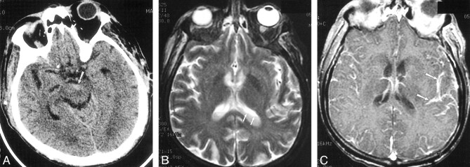
fig 3.
Case 12. 66-year-old patient with diabetes and secondary biliary cirrhosis died following infection with E coli meningitis, diagnosed by C1-C2 puncture. Delayed diagnosis occurred as a result of mild signs and symptoms of infection.
A, Initial CT scan shows debris and slight hyperattenuation (arrow) compared with CSF, producing an irregular level in the interpeduncular cistern.
B, MR imaging was deferred for 2 days because of minor central nervous system symptoms. T2-weighted image (2300/96/2 [TR/TE/excitations]) shows irregular debris (arrows) layering in the lateral ventricles.
C, T1-weighted contrast-enhanced image (500/26/1) shows striking pial subarachnoid space enhancement (arrows), but no ependymal enhancement.

