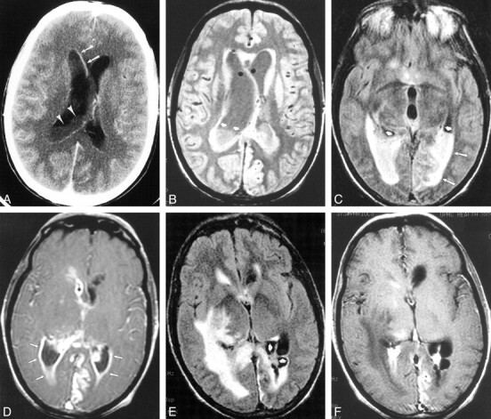
fig 5.

Case 14. 42-year-old patient with diabetes initially presented to an outside facility with meningitis and underwent serial imaging demonstrating the imaging course of ventriculitis during a 7-week period. Streptococcus viridans grew on CSF culture.
A, Initial contrast-enhanced CT scan shows irregular ventricular debris (arrowheads), hydrocephalus, and ependymal enhancement (arrows).
B–D, MR imaging performed 2 weeks later shows ventricular debris (short arrows) and periventricular signal abnormality (long arrows) on proton density–weighted (2000/15/1 [TR/TEeff/excitation]) (B) and FLAIR images (9002/147/1; inversion time, 2200) (C). T1-weighted image (800/20/1) obtained after gadopentetate dimeglumine administration shows extensive ependymal enhancement (arrows) and enhancement of a left posterior cerebral territory infarct.
E and F, There is progression of findings on MR imaging performed 7 weeks after initial presentation. FLAIR (10002/155/1; inversion time, 2200) (E) and T1-weighted images (800/8/1) (F) show loculation of ventricles (arrows) with persistent periventricular signal abnormality, but diminished ependymal enhancement.
