
fig 4.
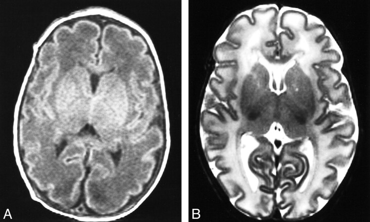
Cortical infolding.
A, Axial T1-weighted image from an infant of 34 weeks' PMA (GA = 34). The frontal and occipital cortices have a similar number of shallow convolutions, and the internal surface of the insula is developing convolutions. The unmyelinated white matter is of low intensity in relation to gray matter. Score: GM2, B1, M3, C3; TMS = 9.
B, Axial T2-weighted image from an infant of 36 weeks' PMA (GA = 36). The frontal and occipital cortices are folded and quite rich in sulci, with deeper sulci evident along the interhemispheric fissure. The insula is more convoluted, and the posterior white matter shows some separation into strands by deeper sulci than in the frontal cortex. Score: GM2, B2, M3, C4; TMS = 11.