fig 1.
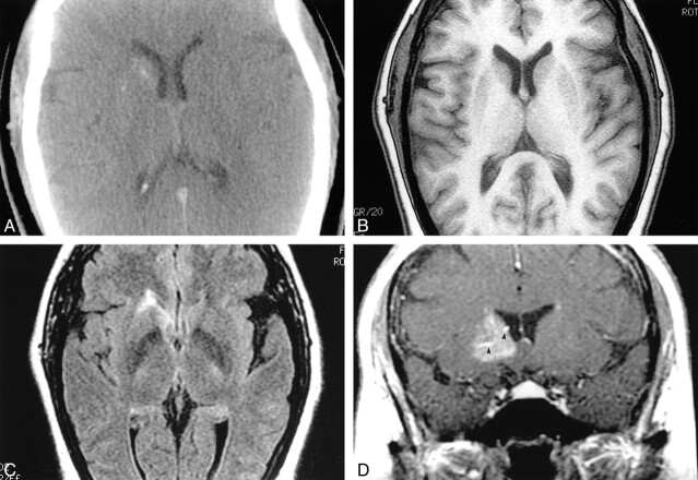
Capillary telangiectasia.
A, Noncontrast CT shows slight hyperattenuation in the right basal ganglia with puntacte calcifications.
B, Noncontrast MR T1-weighted image is normal.
C, Axial FLAIR image (slightly below B) shows high signal intensity in the head of the right caudate nucleus.
D, After gadopentate dimeglumine administration, a coronal T1- weighted image shows diffuse enhancement in the right basal ganglia anteriorly. There is a suggestion of large veins (arrowheads) in the malformation.

