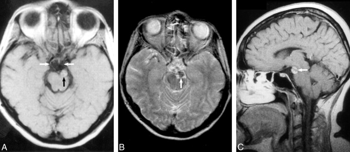
fig 1.
A, Axial T1-weighted unenhanced spin-echo image (800/25/3 [TR/TE/excitations]) demonstrates an extraaxial mass (black arrow) within the interpeduncular cistern that compresses the left cerebral peduncle and displaces the left oculomotor nerve medially (white arrows indicate the oculomotor nerves). The mass contains heterogeneous foci of high signal intensity.
B, Conventional spin-echo axial T2-weighted image (2400/100/2) confirms a heterogeneous intermediate mass with low signal intensity (arrow) inseparable from the terminal basilar and left posterior cerebral arteries.
C, Sagittal enhanced T1-weighted image (550/25/4) following intravenous administration of gadopentetate dimeglumine confirms heterogeneous enhancement (arrow) and normal suprasellar and pineal region cisterns.

