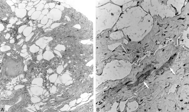
fig 4.
A, Low-power (original magnification ×100) H&E photomicrograph demonstrates dense collagen bundles admixed with fat cells, nerve filaments, and blood vessels.
B, Higher power (original magnification ×400) immunoperoxidase (anti-desmin) preparation reveals scattered skeletal muscle fibers (arrows) interspersed within the dense collagenous tissue, features consistent with teratoma.

