fig 1.
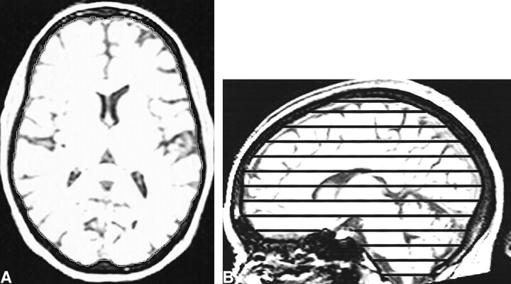
T1-weighted MR images with sagittal and axial views. The intensity windowing is as used for segmentation. The TIV is calculated by summation and linear interpolation of the segmented axial slices.
A, The total intracranial area is shown on one axial section.
B, The axial sections used to sample the total intracranial volume are marked on the sagittal view.

