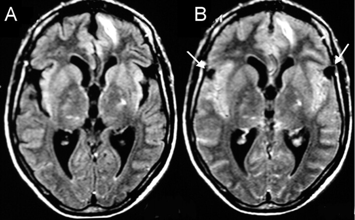
Fig 1.
A 40-year-old man with parenchymal neurocysticercosis and suspected subarachnoid involvement.
A, MR FLAIR image (1.0 T; TR, 11,000 ms; TE, 140 ms; TI, 2600 ms) shows no extraaxial lesions.
B, FLAIR image after 100% O2 for 5 minutes shows two small cysts in the Sylvian fissures confirming racemous neurocysticercosis.

