Fig 1.
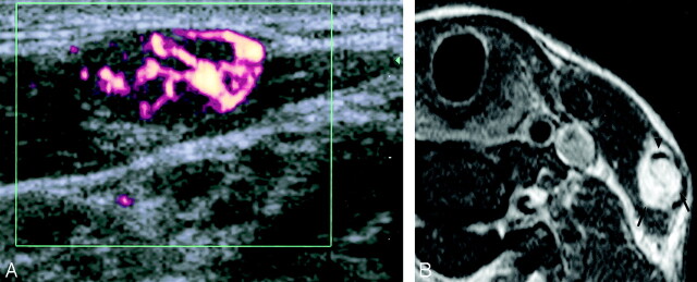
Case 1. Sonographic and MR images obtained in a 31-year-old Indonesian woman with a vascular malformation of the left external jugular vein.
A, Transverse power Doppler sonography shows vascularity within a well-defined, hypoechoic, heterogeneous mass.
B, Axial T1-weighed fat-suppressed contrast-enhanced MR imaging image (TR/TE, 450/15) shows intense enhancement of the mass (arrows) and its relationship to the left external jugular vein (arrowhead).

