Fig 3.
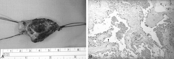
Case 2. Pathologic and histologic sections of a specimen obtained by excision of the left external jugular vein vascular malformation.
A, Gross specimen of the excised lesion shows a well-circumscribed mass with the proximal and distal vascular stumps of the external jugular vein.
B, High-power histologic view of the vascular channels. Note the lining flattened endothelial cells (arrowhead) and the red cells inside the lumens. (Hematoxylin-eosin stain, original magnification, ×120.)

