Fig 4.
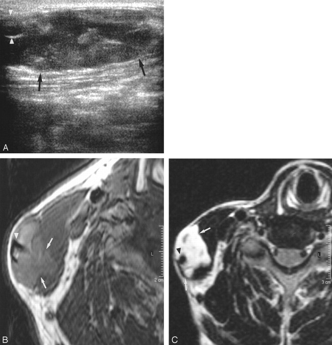
Case 3. Sonographic and MR images obtained in a 37-year-old man with right external jugular vein vascular malformation.
A, Transverse gray-scale sonography shows an ill-defined, hypoechoic, heterogeneous mass (arrows) inseparable from the external jugular vein (arrowheads).
B, Axial T1-weighed spin-echo MR imaging image (TR/TE, 425/18) shows a slightly hyperintense mass (arrows) with ill-defined edges closely related to the right external jugular vein (arrowhead).
C, Axial fat-suppressed T2-weighed MR imaging image (TR/TE, 2500/108) shows a hyperintense mass (arrows) closely related to and inseparable from the right external jugular vein (arrowhead). Note its infiltration into the adjacent sternocleidomastoid muscle.

