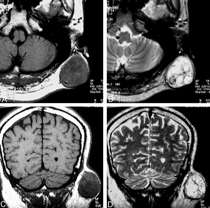
Fig 3.
Axial (A and B) and coronal (C and D) MR images. The mass (4 × 5 cm in diameter) is seated within the subcutaneous adipose tissue. The tumor is heterogeneously hypoisointense and surrounded by a hypointense thin capsule on T1-weighted images (A and C). The multiple, hyperintense nodules are separated by a hypointense, internodular structure on T2-weighted images (B and D).

