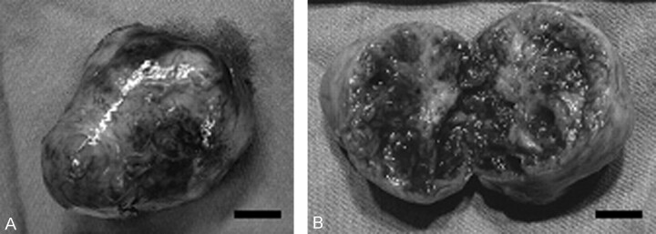
Fig 4.
Gross specimen of the removed tumor. The tumor is covered by smooth fibrous capsule (A) and composed of well-circumscribed, soft to rubbery-firm nodules (B). The cut surface of the tumor is grayish white to yellow and there are scattered macroscopic hemorrhages (B). Scale bars: 1 cm in both A and B.

