
Fig 2.
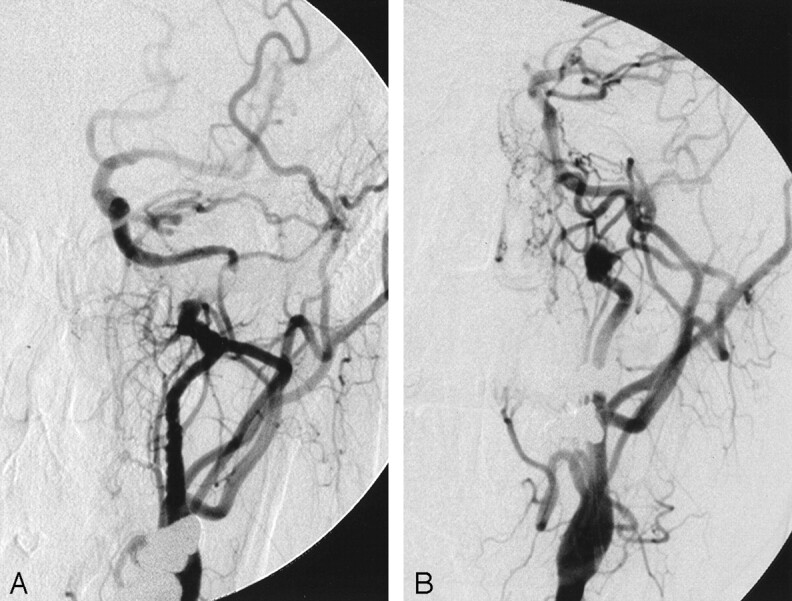
After transfer to consider extracranial-intracranial bypass, a repeat left common carotid angiogram 1 week after initial presentation (A) shows recanalization of the left internal carotid artery with slow flow into the intracranial circulation and dissection of the distal cervical internal carotid artery. A follow-up left common carotid angiogram (B) obtained to confirm a pseudoaneurysm suspected because of CT angiography findings shows a dissecting pseudoaneurysm of the distal cervical internal carotid artery situated on a redundant loop of vessel with an associated significant stenosis.