
Fig 1.
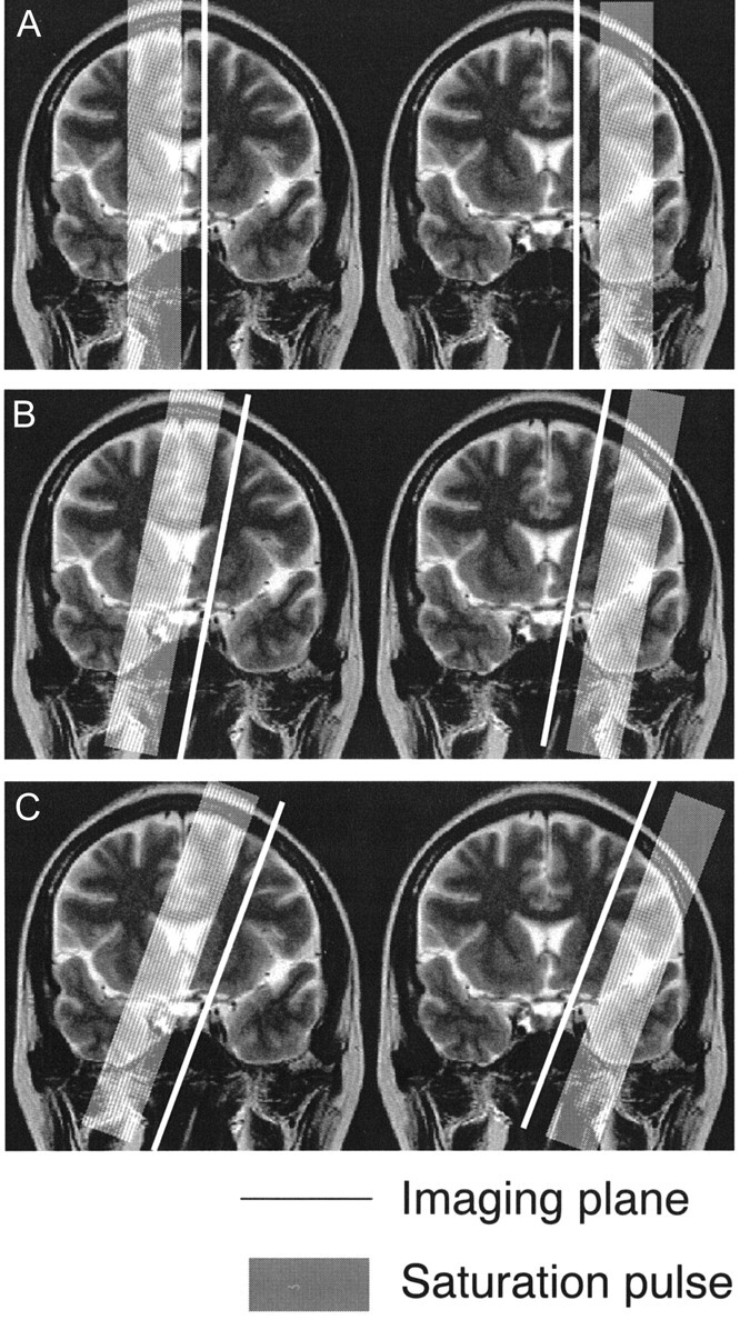
Imaging plane for arterial spin labeling method. We used three imaging planes: plane A, sagittal plane; plane B, 10-degree laterally tilted sagittal plane; plane C, 20-degree laterally tilted sagittal plane. All three imaging planes included the internal carotid artery top. ACA is located on the medial side of the imaging plane, and MCA is located on the lateral side. In the frontal lobe, plane A includes the superior frontal gyrus and plane C includes the middle frontal gyrus. Plane B is located around the superior frontal sulcus in most cases.