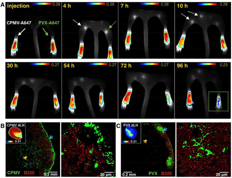
Figure 7:
Draining lymph node (dLN) homing of CPMV and PVX loaded with Alexa Fluor 647 dye. (A) Longitudinal fluorescence imaging of dLN trafficking and retention (dashed arrows) following S.C. injection into the left (CPMV 50μg/20μl) and right (PVX 50μg/20μl) footpads of FVB/N mice: indicating sustained dLN retention of CPMV as compared to PVX. (B) and (C) Confocal microscopy visualization of immunofluorescence staining (green) and imaging of CPMV and PVX accumulations (insets A and B, respectively) in the brachial dLN 12 hours following S.C. injection behind the mice’s neck: showing retention of CPMV in the subcapsular sinus (blue arrow) and T cell zones (yellow arrow), while PVX accumulate (blue arrow) in the follicle zones (red area, anti-B220 antibody staining). Reprinted from [268], Copyright 2016, with permission from Elsevier.

