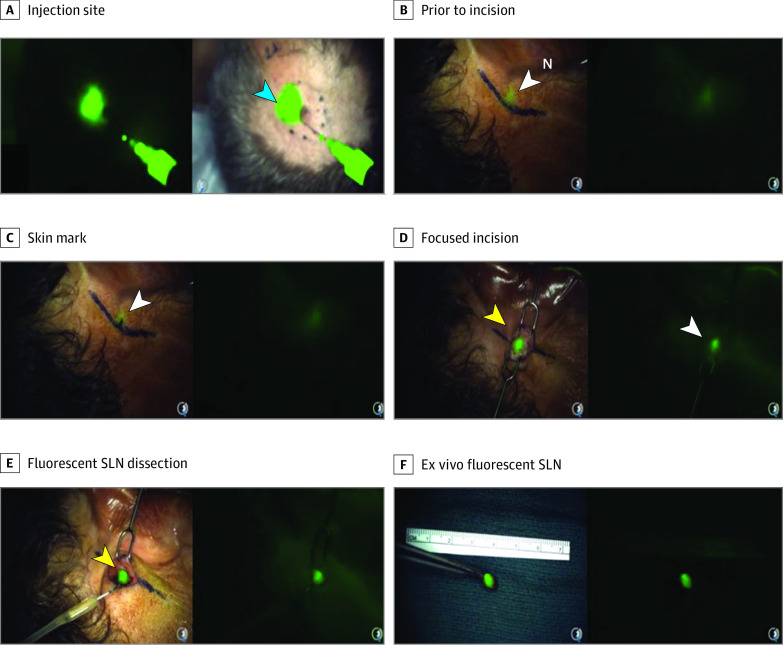
Figure 2. Real-Time Transcutaneous Visualization of a Postauricular Sentinel Lymph Node (SLN) Using Nanoparticles.
A male patient in his 50s with a scalp melanoma was injected peritumorally with integrin-targeting, dye-encapsulated nanoparticles, surface modified with polyethylene glycol chains and cyclic arginine-glycine–aspartic acid–tyrosine peptides (green signal in A). Focal fluorescence was seen through the intact skin overlying a postauricular SLN (B) using the Spectrum camera system (Quest) for real-time optical imaging guidance. Limited extent of surgical dissection (C) and size of the resection cavity (D) relative to the planned area of dissection unaided (ie, marked line in panel C is approximately 3 times larger than that actually drawn on the basis of the particle signal). Images are derived from the intraoperative video (Video).

