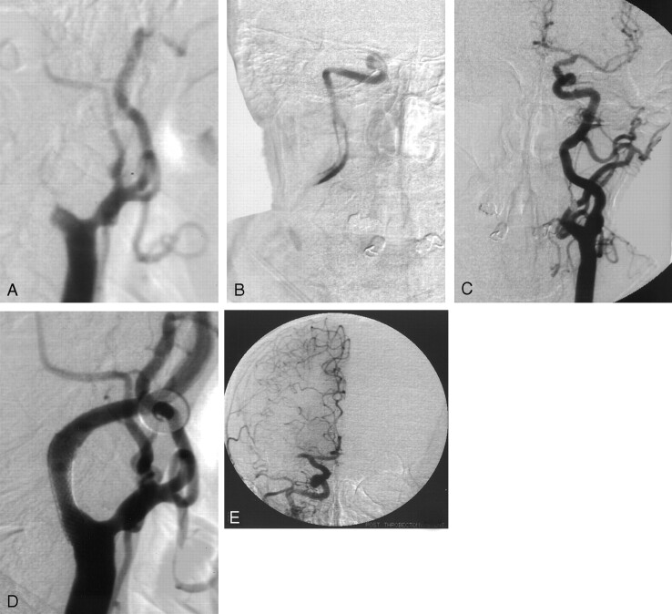
Fig 1.
Case 1. A 67-year-old man presented with resolving left hemiparesis.
A, Baseline right lateral common carotid artery (CCA) angiogram shows near-occlusion of the right ICA with a string sign of slow flow into the proximal ICA.
B, After predilation of the proximal ICA stenosis, this baseline right ICA angiogram reveals the presence of extensive thrombus.
C, Baseline left CCA angiogram reveals some right ACA cross-filling but no right MCA filling.
D, Postprocedural right lateral CCA angiogram shows less than 20% residual ICA stenosis. A Wallstent in the ICA is visible at the carotid bifurcation.
E, Although the right MCA has a persistent mid-M1 occlusion, the postprocedural right CCA angiogram shows good distal filling of the MCA branches via pial collaterals from the ACA.

