Fig 2.
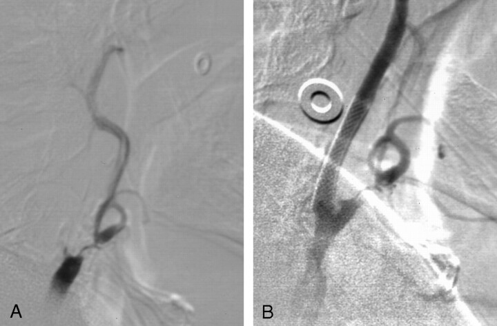
Case 2. A 72-year-old man had global aphasia and right hemiparesis.
A, Baseline left lateral CCA angiogram shows complete occlusion of the cervical ICA. Flow through a 50% stenosis of the external carotid artery remains visible.
B, Postprocedural left lateral CCA angiogram demonstrates essentially complete resolution of the ICA occlusion. The Wallstent placed in the left ICA is visible at the carotid bifurcation.

