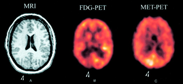
Fig 1.
Images from the case of a 34-year-old woman who presented with medically intractable epilepsy of complex partial and left visual seizures.
A, Contrast-enhanced MR image (MRI) of the brain shows thickening within the right occipital lobe and in the posterior aspect of the right temporal lobe, suggestive of polymicrogyria/pachygyria.
B, [18F]Fluorodeoxyglucose positron emission tomographic scan (FDG-PET) of the brain shows focal area of decreased glucose uptake in the right occipital lobe.
C, [11C]Methionine positron emission tomographic scan (MET-PET) of the brain shows enhanced methionine uptake in the right occipital lobe.

