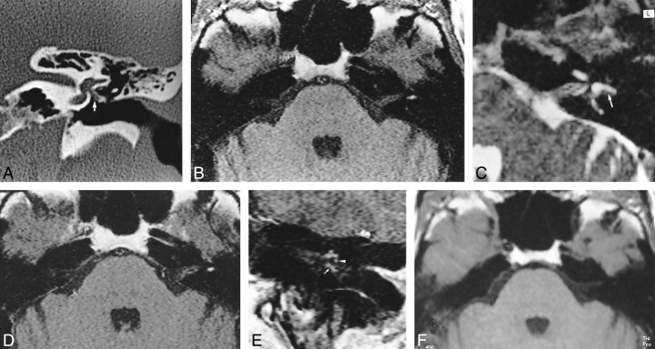
fig 1.
58 year-old woman with postoperative hemorrhagic labyrinthitis 2 months after left-sided stapedectomy.
A, Coronal CT scan of left temporal bone shows a hypodensity along a well-located prosthesis (arrow). The lateral semicircular canal is normal.
B, Axial T1-weighted MR image shows abnormal signal intensity of the left lateral semicircular canal, as compared with right ear.
C, Axial T2-weighted MR image (2500/90) shows normal hyperintensity of the lateral semicircular canal (arrow).
D, Contrast-enhanced axial T1-weighted MR image (500/15) shows no change in the signal intensity of the labyrinth.
E, Contrast-enhanced coronal T1-weighted MR image shows an enhancing mass engulfing the prosthesis (arrow). Note the normal enhancement of the tympanic portion of the facial nerve (arrowhead) under the abnormal lateral semicircular canal.
F, Follow-up axial T1-weighted MR image at 1 year shows disappearance of the high signal intensity of the lateral semicircular canal.

